
“X-Ray Vision” for Surgeons: AI Holograms Transform Cancer Surgery
Editor’s note:Among the 2026 China Integrated Oncology Diagnosis and Treatment Technology Guidelines series, the Holographic Imaging Technology Guidelines stand out as the first of their kind globally. By integrating artificial intelligence, this technology converts conventional two-dimensional CT, MRI, and other imaging data into three-dimensional digital holograms. It is transforming preoperative planning, intraoperative navigation, medical education, and patient communication, particularly in urologic oncology surgery.
Professor Zhu Gang [Note: Prof. Zhu serves as Associate Chief Medical Officer, Chair of Surgery & Chair of Urology at United Family Healthcare Beijing], lead editor of the guidelines and Chair of the Integrated Rehabilitation Committee for Genitourinary Oncology at the China Anti-Cancer Association, recently gave an exclusive interview to Oncology Frontier. In the discussion, he explained how AI-powered holographic imaging works, shared clinical applications, outlined future directions, and discussed the evolving role of urologic surgeons within an integrated care framework. An edited transcript is provided below.
The Role of Holographic Imaging in Preserving Organ Function in Urologic Tumor Surgery
Q1
Professor Zhu, in your presentation, you highlighted the use of AI-powered holographic imaging to help preserve organ function in urologic tumor surgery. How does this technology work, and what advantages does it offer over traditional 2D imaging?
Today I presented on the application of holographic imaging technology in organ-preserving surgery for urologic tumors. Public understanding of holography often comes from optical projections, like the three-dimensional images seen in science fiction.
With advances in artificial intelligence, however, we can now generate digital holograms directly from medical imaging data, without the need for complex optical systems. In clinical practice, this means converting standard 2D CT, MRI, or even ultrasound images into interactive 3D models.
Human anatomy is inherently three-dimensional, yet most medical imaging is presented in two dimensions. While clinicians are trained to mentally reconstruct these images, this is difficult for patients. Holographic imaging bridges this gap by providing an intuitive, manipulable 3D view.
The model can be rotated and examined from any angle, clearly showing the relationship between the tumor and surrounding structures such as blood vessels and collecting systems. This improves patient understanding and supports more effective communication around surgical planning.
The technology also has significant value in medical education.
Traditionally, anatomical learning relied heavily on cadaver dissection. Now, full-body imaging data can be converted into detailed 3D holograms, allowing students to visualize anatomical relationships more clearly and dynamically.
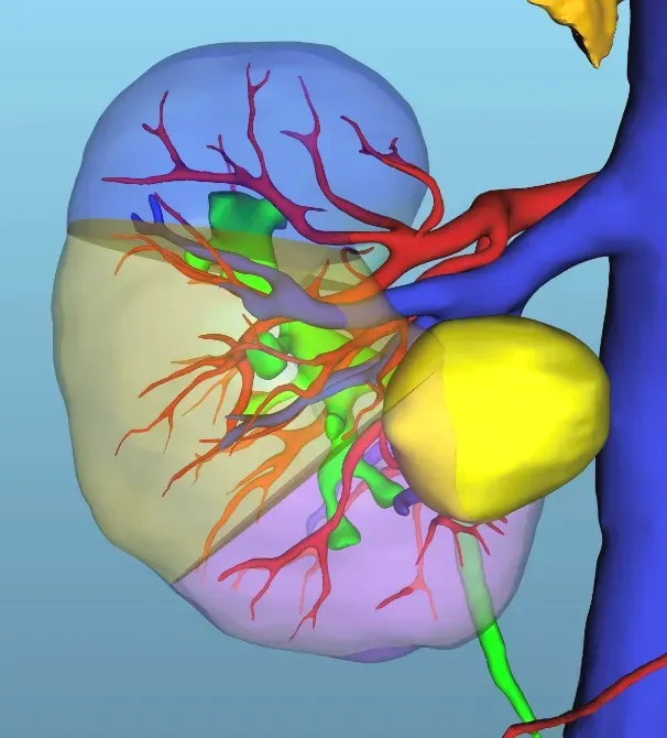
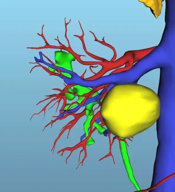
Clinically, holographic imaging is most widely used in preoperative planning. Surgeons can enlarge and manipulate the model to design precise surgical approaches. During surgery, it can also support intraoperative navigation by overlaying holographic data onto endoscopic views, helping guide more accurate and controlled procedures.
The China Anti-Cancer Association has now formally incorporated this technology into its guidelines. Our multidisciplinary team, including experts from urology, hepatobiliary and pancreatic surgery, thoracic surgery, and other fields—worked together to standardize its application. Ultimately, holographic imaging enhances surgical precision and helps maximize organ function preservation.
Q2
Can you share a clinical case where this technology helped preserve organ function?
The value of any new technology lies in its clinical impact. I have been using holographic imaging since 2017, and its benefits have become increasingly clear, particularly in kidney and adrenal tumors.
For kidney tumors, a detailed anatomical understanding is critical. The kidneys have a complex vascular structure, and damage to major vessels can lead to severe complications. Holographic imaging allows for precise preoperative mapping, enabling surgeons to preserve as much healthy renal tissue as possible.
Not all tumors are superficial; some are located deep within the renal parenchyma or near the renal hilum, where critical vessels and the ureter converge. In these cases, holographic visualization clearly shows the tumor’s spatial relationship with surrounding structures, allowing for careful surgical planning that balances complete tumor removal with maximal tissue preservation.
In adrenal tumors, the challenge is often localization. Some functional tumors are extremely small, for example, aldosterone-producing adenomas. We once treated a tumor measuring just 6 mm. Using holographic imaging in combination with intraoperative ultrasound, we were able to localize and completely remove the lesion, resulting in a cure.
Overall, holographic imaging has proven highly valuable in clinical practice. With the release of these guidelines, I expect its adoption to expand, benefiting more patients.
Q3
As lead editor of the world’s first Holographic Imaging Technology Guidelines, how do you see this technology evolving?
This is a key question. Similar technologies have been described using terms like “3D reconstruction” or “augmented reality.” By standardizing the term “holographic imaging technology,” the China Anti-Cancer Association has taken an important step in reducing confusion and promoting broader adoption.
Although these technologies are used internationally, this is the first guideline to clearly define and standardize the field. Our team included experts from multiple surgical disciplines, reflecting the wide applicability of this approach.
I am very optimistic about its future. Holographic imaging will play an increasingly important role in oncologic surgery, particularly in improving precision and preserving organ function.

Q4
From a clinical needs perspective, what improvements could be made to holographic imaging, and what problems do you hope to solve in the next phase?
While promising, the technology is not without limitations.
Currently, generating holograms often requires sending imaging data to external companies, which raises concerns about patient privacy. A key development direction is enabling in-hospital deployment so data can be processed securely within radiology departments.
Another challenge is real-time intraoperative navigation. Surgery is dynamic, organs move and deform during procedures. While static overlays are feasible, achieving accurate real-time alignment between holograms and surgical fields remains technically complex. That said, progress is being made, and I believe real-time navigation will be achievable in the near future.
Q5
What barriers exist to broader adoption?
Clinical validation is essential. We need robust evidence, including multicenter studies, to confirm effectiveness and accuracy.
Cost is another important factor, particularly for wider adoption. As usage increases and AI reduces reliance on manual processes, costs are expected to decline.
At the same time, we aim to expand both international collaboration and grassroots implementation. With continued research, innovation, and knowledge exchange, this technology has strong potential for global adoption.
Q6
How has integrated medicine influenced your role as a urologic surgeon?
The role of the surgeon has evolved. We are no longer focused solely on performing procedures, but on managing the entire patient journey, from diagnosis through treatment and follow-up.
This includes evaluating the need for neoadjuvant therapies, planning intraoperative strategies, and coordinating postoperative treatments such as chemotherapy, radiotherapy, or immunotherapy. Long-term follow-up is also critical to monitor for recurrence or metastasis.
A comprehensive, patient-centered approach is essential to achieving the best outcomes.
Q7
Why is holistic thinking increasingly important in specialized medicine?
Cancer care requires an integrated approach. The full continuum—from prevention and screening to diagnosis, treatment, and rehabilitation—cannot be managed by a single specialty.
Effective care depends on collaboration among surgeons, oncologists, radiologists, pathologists, and allied health professionals, including nursing, nutrition, and psychological support. This multidisciplinary model ensures more comprehensive and coordinated care.
Q8
What practical benefits does this approach bring?
An integrated, holistic approach improves both clinical outcomes and patient experience. By aligning care across all stages of the disease and across specialties, we can deliver more effective treatment and help patients achieve longer survival and better quality of life.